Monday, November 28, 2016
A "Good Enough" Stage Micrometer
http://www.microscopy-uk.org.uk/mag/indexmag.html?http://www.microscopy-uk.org.uk/mag/artoct02/hwmicrometer.html
Sunday, November 27, 2016
Life cycles algae & plants
Life cycles algae & plants
|
|||||
Organisms, from little moss to
man, go during their life through a number of biological phases, generation
after generation. The following "biological phases" are encountered
during the life cycle: the formation of gametes (the
sexual reproductive cells), the fusion of male and female gametes (~ fertilization)
to a zygote, and a period of growth and development (cell
differentiation and morphogenesis) that occurs at various time sequences,
depending on the phylogenetic group to which the organism belongs. Within the
life cycle, meiotic divisions (more) take place, in
which cell proceed from a diploid to a haploid stage;
in the diploid stage two homologous copies of each chromosome are present in
the cells and in the haploid stage only one sample of each chromosome in
present in each cell. Gametes are always haploid, whereas zygotes are
diploid. Furthermore, during the life cycle mitotic divisions occur
(more about
mitosis). Depending on the phylum to which the organism belongs, these
mitotic divisions occur in the haploid, or diploid phase, or in both the
haploid and diploid stages. More here about here below.
In these web modules one finds descriptions of the life cycles of brown, red and green algae and that of various phyla of land plants. The characteristics are shown according to typical representants of each group, the same as those that are discussed in the course "Evolution and Development of the Plant" (Radboud University Nijmegen). The classification and terminology in these webpages follows the handbook that is used during lectures and practical courses: "Biology, Campbell, Reece, Urry, Cain, Wasserman, Minorsky en Jackson, 2008. Pearson International Edition (eds).San Francisco. 1267 pp". 
Three types of life cycles can be distinguished on account of the timing of
the mitoses, in the haploid and/or diploid phase:

Colofon
This series webpages on the life
cycle in plants is based on course notes and other teaching material of Mieke
Wolters-Arts, Celestina Mariani, Jan Derksen, the late G. van den Ende and
Luud J.W. Gilissen. Dr Gerard van den Ende had developed an extended manual
enriched with numerous drawings of life cycles, which went along with the
practical courses that he taught. The microscopical work and the layout are
by Liesbeth Pierson (contact), Wilbert Janssen and Ines Schulten.
The web structure was developed by Remco Aalbers.
|
|||||
Friday, November 25, 2016
Inoculation of 50ml growth culture take from petri dish cultures into 500ml growth media.
Inoculation of 50 ml growth culture take from petri dish cultures into 500 ml growth media.

After 5 days growth of media taken from the front yard growth media petri dish sample and inoculate a 500 ml growth media.
Before this I examined the sample under microscope and found nothing but what I am labeling Characium pringsheimii based upon visual comparison.
very distinctive structure


After 5 days growth of media taken from the front yard growth media petri dish sample and inoculate a 500 ml growth media.
Before this I examined the sample under microscope and found nothing but what I am labeling Characium pringsheimii based upon visual comparison.
very distinctive structure


More Characium pringsheimii from FY11-01-2016
I was able to get very good detail on what I am thinking is Characium pringsheimii.
I also found a couple of cells that had apparently ruptured and captured a small video of parasites feeding on the expelled inards.


I also found a couple of cells that had apparently ruptured and captured a small video of parasites feeding on the expelled inards.


Ankistrodesmus acicularis for Percy Priest Lake
I now have significant growth in the Percy Priest Lake algae sample sample taken at the dam.
I examined the specimen under microscope and found several species of algae in the sample, including Ankistrodesmus acicularis.
Here are some videos and images taken 11-22-2016
Immobile Ankistrodesmus


Taken 11-25-2016 - showing more Characium and possible Euglena - doesn't look like Ankistrodesmus


I examined the specimen under microscope and found several species of algae in the sample, including Ankistrodesmus acicularis.
Here are some videos and images taken 11-22-2016
Immobile Ankistrodesmus


Monday, November 21, 2016
FY11012016 Front Yard Algae Culture Upscale
Today I went ahead and upscale the FY11012016 Front Yard Algae Culture from 100ml to 2 liter using my standard MiracleGro media to see how well it upscales.


11/21/2016

12-03-2016 - ready to harvest??

Sunday, November 20, 2016
We have algae culture growth in petri dish!!!
I went on a 3 day job and came back and found that I had algae culture growth in my petri dish simply placed in my newly created bioreactor.
AGAIN, the algae from the front yard pond grew successfully on the miraclegro algae agar from the recipe in my previous post from Doane College Algae Lab YouTube videos.
Here is a great link to all of their pertinent video series. Awesome resource!!
http://doanealgae.wixsite.com/algaegrowingresource/videos
And now the images of my petri dishes!! MESSY MESSY.


Being overly excited to cultivate a single species I have created 3 50 ml glass culture bottles made from Cracker Barrel maple syrup bottles!! I took small 'single colony' scrapping from 3 seperate areas and added to the bottles.
Here is day one images -

I placed the corresponding petri dishes behind the corresponding culture. 11-20-2016

Here is an image from 5 days ago - 11-15-2016. (significant difference in growth!!

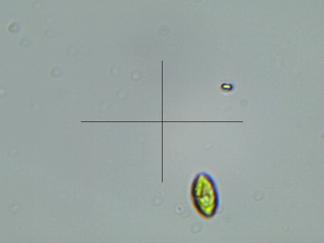
I have Characium pringsheimii - i think
After observing my algae from the front pond, I dont think I have Euglena. I may have observed one or two but the large majority of the sample is more characteristic of Characium pringsheimii - http://protist.i.hosei.ac.jp/pdb/images/Chlorophyta/Characium/pringsheimii_2.html
Genus: Unicellular; cell body ellipsoidal or spindle-shaped, attached on a substrate at one end; a single chloroplast plate-like in shape, with a pyrenoid (Guide book to the "Photomicrographs of the Freshwater Algae", 1998).
Their image -


My microscope video
Genus: Unicellular; cell body ellipsoidal or spindle-shaped, attached on a substrate at one end; a single chloroplast plate-like in shape, with a pyrenoid (Guide book to the "Photomicrographs of the Freshwater Algae", 1998).
Their image -

Secondary resource - http://www.plingfactory.de/Science/Atlas/Kennkarten%20Algen/01_e-algae/Chlorophyta/e-source/Characium%20sp.1.html
Their images -


Tuesday, November 15, 2016
OMG Euglena eat algae!!! OH HAYUL NAW!!!
http://www2.fcps.edu/islandcreekes/ecology/euglena.htm
Euglena gracilis and other euglena are green because they eat green algae. They keep the algae inside their bodies and use it to make their own food. These green parts inside the Euglena's body are called chloroplasts.
SO I HAVE IDENTIFIED AN ALGAE EATER IN MY ALGAE!!!
OH HELL NAW!!!
Euglena gracilis and other euglena are green because they eat green algae. They keep the algae inside their bodies and use it to make their own food. These green parts inside the Euglena's body are called chloroplasts.
SO I HAVE IDENTIFIED AN ALGAE EATER IN MY ALGAE!!!
OH HELL NAW!!!
Using Microscope Attempt to identify algae species
Attempt to identify algae species using - - http://www.landcareresearch.co.nz/resources/identification/algae/identification-guide/identify/guide
Here are reference images of my summer algae sample that shows no sign of dying after 5 months.


And some videos


These videos show this species changing shape from circular to spindle and are not algae but PROTISTS!! - After observing with the 25x eyepiece instead of the usb camera - I noticed that there is definitely a split end that I can seen vibrating - meaning it HAS FLAGELLA.
Wikipedia says - https://en.wikipedia.org/wiki/Euglenid this about Euglena.

So at this stage I am waiting to see if the two samples I have in petri dishes come to fruition ...........then isolate and culture...JUST ALGAE, NO PROTISTS ALOWED!!!!
Here are reference images of my summer algae sample that shows no sign of dying after 5 months.


And some videos
Show the dominate algae species is definately spindle shaped like this -

Single cellular with non distorting walls and no visible flagella.
My observation to answers to the identification guide
- Algae are unicellular - not joined to neighbouring cells
- Cell wall is smooth, cell outline round to ellipsoidal, and featureless
- Cells containing membrane-bound organelles, such as chloroplasts and nuclei.
- Cell with rigid cell wall, lacking shallow groove at equator, and motile, propelled by one or more waving hair-like structures (flagella) - EXCEPT I CAN NOT SEE FLAGELLA, my microscope may just not be good enough to resolve - but had to say this to get to spindle shape..
- Unicellular chlorophyte with an elongated, usually spindle-shaped cell with two short flagella emerging from one end. There is a single chloroplast that may contain a visible eyespot. If flagella are not visible, the cells could easily be confused with the spindle-shaped cells of other genera.
Chlorogonium (Chlamydomonadaceae)
looks almost identical -

But NO FLAGELLA and the shape isn't exactly like mine -
nothing on youtube on Chlorogonium.
sample is closer to - Euglena (Euglenidae) - http://www.landcareresearch.co.nz/resources/identification/algae/identification-guide/identify/guide/unicellular/euglena

You tube videos on Euglena viridis - much higher resolution than I can get. AT THIS TIME.
I wish I could afford a microscope as good as these!! DAMN!!
This video was recorded 400x!! I have to goto 650x just to see them. Meaning these appear to be alot larger!!
These videos show this species changing shape from circular to spindle and are not algae but PROTISTS!! - After observing with the 25x eyepiece instead of the usb camera - I noticed that there is definitely a split end that I can seen vibrating - meaning it HAS FLAGELLA.
Wikipedia says - https://en.wikipedia.org/wiki/Euglenid this about Euglena.

So at this stage I am waiting to see if the two samples I have in petri dishes come to fruition ...........then isolate and culture...JUST ALGAE, NO PROTISTS ALOWED!!!!
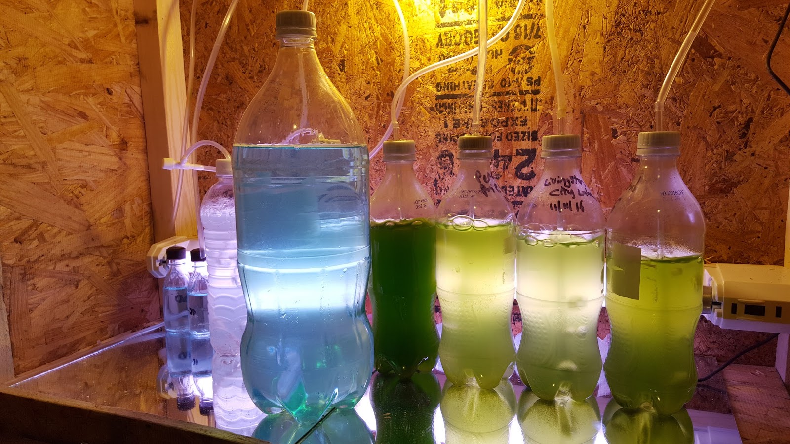
My Simple Square Box PhotoBioReactor PBR Design using recycled soda bottles
In an earlier post I showed my first PBR design from 2X4x and a leaning vertical tube design. While this design worked well. It was a real pain to detach and reattach the hoses at the bottom of the tube to clean or empty their contents.

Then I designed a semi-circular PBR using an instructable and alot of guess work to build a semicircular shaped pbr and while it made removing and installing the tubes a little bit easier it was still too cumbersome and gangly to work with. Setting the lighting and air pump timers was a bitch. Also I dont think the tubes were close enough to the grow light.


THE main thing I was having trouble with was using the vertical tubes, each time.
The last straw with the tubes was when I noticed that despite the air stones at the bottom of the tubes I was getting SIGNIFICANT setting below the air stones at the bottom of the tubes.

Again not very practical. It was fun to build but just sucked.
I found that every time I took a sample it was in a 20 oz soda bottle and for some reason just adding a 100 ml of miracle gro algae media I created earlier the algae grew better than in the tubes.
So I decided to eliminate the tubes and just use recycled soda bottles and design a simple easy to change and clean and move things around in pbr. I know a run on sentence.
Here is the first square design made from nothing more than 2x4s - oh I had to buy the plexiglass but the 2x4s I alread had, from the previous leanto design.


This design was WAY easier to use and more practical. I decided to try putting the light underneath.
This didnt provide enough light so I redesigned it to be enclosed ( slightly more aesthetically pleasing - and lighting from the side of the bottles for better light penetration.
Here is what I came up with - definitely not my final design - designing is the fun part!!
All from a 4'X4' sheet of osb, I had it laying around.

screwed together with 2x2s

Placed the air pump and timers on top for easy access.


Two 4 gang controls in back.
Video of prettier design in operation - I used mirror tiles to try reflect light back up towards samples.
So the dark green sample, Second from the left is the sample I have been keeping going every since summer. I am still yet to get anything to grow inside my incubator so I have also placed 2 more samples of my summer sample to the far left.
TIME TO USE MY MICROSCOPE TO IDENTIFY WHICH ALGAE SPECIES ARE IN THE SAMPLE, AND WHICH IS THE DOMINANT. BUT I REALLY WANT MY COLONIES TO GROW SO I CAN ISOLATE ...........

Then I designed a semi-circular PBR using an instructable and alot of guess work to build a semicircular shaped pbr and while it made removing and installing the tubes a little bit easier it was still too cumbersome and gangly to work with. Setting the lighting and air pump timers was a bitch. Also I dont think the tubes were close enough to the grow light.
Here are a couple of images for reference.


THE main thing I was having trouble with was using the vertical tubes, each time.
The last straw with the tubes was when I noticed that despite the air stones at the bottom of the tubes I was getting SIGNIFICANT setting below the air stones at the bottom of the tubes.

Again not very practical. It was fun to build but just sucked.
I found that every time I took a sample it was in a 20 oz soda bottle and for some reason just adding a 100 ml of miracle gro algae media I created earlier the algae grew better than in the tubes.
So I decided to eliminate the tubes and just use recycled soda bottles and design a simple easy to change and clean and move things around in pbr. I know a run on sentence.
Here is the first square design made from nothing more than 2x4s - oh I had to buy the plexiglass but the 2x4s I alread had, from the previous leanto design.


This design was WAY easier to use and more practical. I decided to try putting the light underneath.
This didnt provide enough light so I redesigned it to be enclosed ( slightly more aesthetically pleasing - and lighting from the side of the bottles for better light penetration.
Here is what I came up with - definitely not my final design - designing is the fun part!!
All from a 4'X4' sheet of osb, I had it laying around.

screwed together with 2x2s

Placed the air pump and timers on top for easy access.


Two 4 gang controls in back.
Video of prettier design in operation - I used mirror tiles to try reflect light back up towards samples.
So the dark green sample, Second from the left is the sample I have been keeping going every since summer. I am still yet to get anything to grow inside my incubator so I have also placed 2 more samples of my summer sample to the far left.
TIME TO USE MY MICROSCOPE TO IDENTIFY WHICH ALGAE SPECIES ARE IN THE SAMPLE, AND WHICH IS THE DOMINANT. BUT I REALLY WANT MY COLONIES TO GROW SO I CAN ISOLATE ...........
Subscribe to:
Comments (Atom)